Pododermatitis in dogs: leading causes

Introduction
Pododermatitis is skin inflammation that can affect interdigital spaces, paw pads, nail beds and adjacent tissues, and nails.1 The incidence of pododermatitis is the same regardless of age or sex.
However, some breeds have a greater predisposition, and this includes both short-haired (Boxer, Bulldog, Bull Terrier, Pointer) and long-haired breeds (German Shepherd, Golden Retriever, Irish Setter).
2 Affected tissues usually present varying degrees of pruritus, erythema, oedema, comedones, paronychia and nodules which may ulcerate with serosanguineous drainage or a purulent exudate.1,2 The various diseases that cause pododermatitis in dogs have very similar clinical manifestations, so it is impossible to reach an accurate diagnosis based solely on the appearance of the lesions.3
Pododermatitis in dogs: aetiology
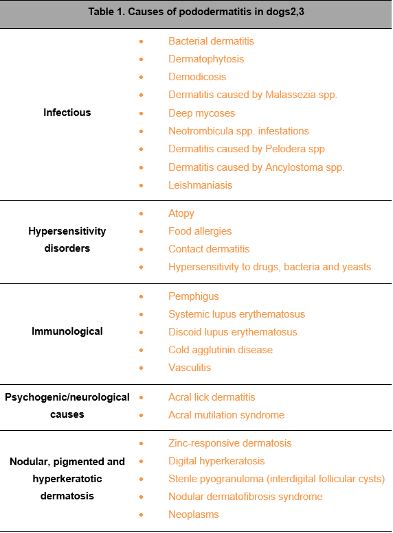
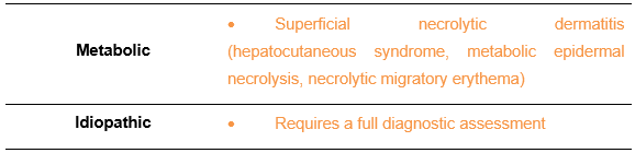
Depending on its aetiology, pododermatitis in dogs can be classified into eight main groups:2.3 (Table 1).
DEMODICOSIS
Many dogs with pododermatitis secondary to demodicosis are misdiagnosed as atopic, so the treatment they receive is ineffective or aggravates the animal’s clinical picture presented.3 Therefore, it is important to bear in mind that dogs with pododermatitis caused by demodicosis do not present pruritus, or it is only mild, but rather they develop nodules and fistulas with a bloody fluid.
ATOPY
Pruritus (which causes intense licking of the feet) is a classic symptom of atopy. Erythema and varying degrees of lichenification and excoriations also occur in atopic pododermatitis.1,3 Furthermore, atopic patients with pododermatitis are often infested withMalassezia spp.
DERMATOPHYTOSIS
Dermatophytes can cause pododermatitis in dogs and, as does demodicosis, they can produce lesions in other areas. However, unlike demodicosis, it rarely courses with nodules with a bloody drainage because dermatophytes live in the most superficial layer of stratum corneum, unlike Demodex spp, which live in deeper layers.3 The most common dermatophytes in dogs are Trichophyton spp., which do neither fluoresce under a Wood’s lamp nor produce arthrospores in the patient’s hair. Consequently, the diagnosis is usually based on a fungal culture. 3

DEEP PYODERMA
Deep pyoderma on the legs is characterised by recurrent nodules, fistulas and drainage channels, similar to those that develop in demodicosis or interdigital follicular cysts (although these generally affect a single interdigital space, usually between toes 4 and 5, while pyoderma tends to affect several). Demodicosis is sometimes complicated by the coexistence of deep pyoderma. As such, when treating a dog with these signs, start by searching for Demodex spp. mites and if the result is negative, carry out a culture and antibiogram if deep pyoderma is suspected.3
IDIOPATHIC PODODERMATITIS
Some cases of pododermatitis diagnosed as idiopathic following a full assessment respond to immunomodulatory therapy, which suggests an immune-mediated aetiology in these patients, but this observation has not been well documented.2
Diagnosis
- In addition to a complete physical examination and a thorough medical history, the first steps should include amicroscopic examination of the hair, deep skin scraping and cytology.
- If there is a suspicion or evidence of dermatophytes or bacteria, perform the corresponding culture and antibiogram, where appropriate.
- If this initial approach does not lead to an accurate diagnosis, the next step is to consider a skin biopsy.
- In addition, thyroid or adrenal function should be tested in patients with systemic signs and imaging tests are recommended if a foreign body is suspected.1,2
Treatment
Pododermatitis in dogs is often self-perpetuating, multifactorial and resistant to empirical treatment, which makes it a frustrating disease, for both the veterinary surgeon and owners.1 Consequently, a good diagnostic protocol that can establish and treat the primary cause is essential.
Conclusions
Pododermatitis in dogs cannot be considered a definitive diagnosis, but rather a clinical manifestation of a primary disease. Effective management therefore requires an appropriate diagnostic protocol to establish and treat the primary cause of pododermatitis.